Search
SYTO 82 Nuclear Staining Protocol

Nuclear stain for eukaryotic and prokaryotic cells
SYTO 82 is a cell-permeant, non-exclusive nucleic acid stain that shows a large fluorescence enhancement upon binding. In both live and dead eukaryotic cells, SYTO 82 generally shows cytoplasmic or mitochondrial as well as nuclear staining. In addition, SYTO 82 will stain most live and permeabilized bacteria.
This protocol can be used for:
- Nucleic acid (nuclear) staining in fluorescence microscopy
This protocol should not be used for:
- Flow cytometry
You will need the following for this protocol:
- Cells growing in culture
- SYTO 82 Orange Fluorescent Nucleic Acid Stain (Cat. No. S11363)
- Fluorescence microscope
Protocol
Spectral information and storage
| SYTO 82 | |
|---|---|
| Excitation/Emission (nm) | 541/560 |
| Standard filter set | TRITC |
| EVOS Light Cube | RFP |
| Storage conditions | ≤–20°C |
Protocol tips
- Warm to room temperature and briefly centrifuge the DMSO solution to the bottom of the vial each time before use.
- Try multiple dye concentrations in the range from 100 nM to 5 µM to determine the optimal concentration.
- In general, the best results are obtained in buffers that do not contain phosphate, such as Hank’s Balanced Salt Solution (Cat. No. 14025092).
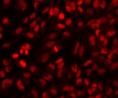
Cells stained with SYTO 82 dye and imaged with the Thermo Scientific CellInsight High-Content System.